


A new technology has been tested for the first time on people which can give a 3-D image of the patient’s arteries and better treat heart diseases. The technology is in the early stages of testing according to a feasibility study published in Circulation: Cardiovascular Interventions, an American Heart Association journal. According to the cardiologists this is a very exciting technology that holds great promise. The study allows doctors to assess more accurately and rapidly the length, branching pattern and angles of heart arteries, as well as any blockages. Cardiologists currently use two-dimensional X-ray images shot from different angles to visualize arteries inside the body. They also inject contrast dye into a thin tube — a catheter — inserted into a patient’s leg artery and threaded up to the heart to produce shadow images during a cardiac catheterization procedure. Although it uses existing X-ray systems, the new software reduces the need for several of the images, thus reducing patients’ exposure to radiation and contrast dye while also decreasing the time doctors need to analyze the images. The comparison of 2-D and 3-D computer images of the coronary artery systems will be tested in multiple centres around the world. It will be formally tested to see the impact on clinical care. Cardiovascular disease is the number one killer in the United States. According to the World Health Organisation, it is responsible for 17 million deaths worldwide. Be a part of Elets Collaborative Initiatives. Join Us for
Upcoming Events and explore business opportunities. Like us on
Facebook , connect with us on
LinkedIn and follow us on
Twitter ,
Instagram. "Exciting news! Elets technomedia is now on WhatsApp Channels Subscribe today by clicking the link and stay updated with the latest insights!" Click here!
 Like
Like
 Dislike


Related Research
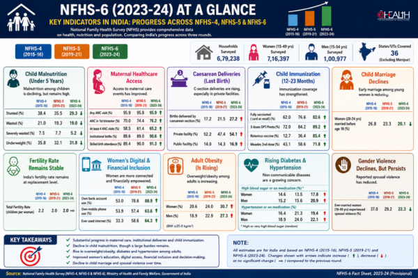
NFHS-6 Reveals India’s Mixed Health Transition: Child Malnutrition Falls, Healthcare Access Improves, But Obesity & Lifestyle Diseases Surge
India’s latest National Family Health Survey (NFHS-6) paints a complex portrait of the country’s public health journ...
By eHealth Network
![]() 30-05-2026
30-05-2026
P&G Health and IMA Unveil India’s First Evidence-Based Patient Recovery Guidelines Focused on Micronutrients
Strengthening post-illness care in India, Procter & Gamble Health Limited has partnered with the Indian Medical Asso...
By eHealth Network
![]() 25-03-2026
25-03-2026

30 Years of Scientific Rigour: DiagnoSearch Reinforces Its Role in Accelerating Pharma Pipelines
DiagnoSearch Life Sciences, one of India’s longest-standing and most trusted Clinical Research Organisations (CROs), h...
By eHealth Network
![]() 03-12-2025
03-12-2025
Servier India Partners with MedGenome & Strand Life Sciences, Expanding AML & CCA Testing
To advance India's precision oncology and equitable access to molecular diagnostics, Servier India, a subsidiary of the ...
By eHealth Network
![]() 11-11-2025
11-11-2025

One in Four Indians at Risk of Heart Disease as Lipid Imbalances Rise Across Age Groups: Metropolis Study | World Heart Day Special
Metropolis Healthcare Limited, one of India’s largest and most respected pathology networks, has released a nationwide...
By eHealth Network
![]() 29-09-2025
29-09-2025

IIT Roorkee Scientists Unveil Novel Drug Candidate to Tackle Antibiotic Resistance Crisis
Scientists at the Indian Institute of Technology (IIT) Roorkee have identified a potential drug candidate capable of res...
By eHealth Network
![]() 12-08-2025
12-08-2025

82% Prefer Diagnostic Centres Over Hospitals in India: ekincare Report
A new data-backed report by ekincare uncovers a decisive change in India’s preventive healthcare landscape, placing di...
By eHealth Network
![]() 09-07-2025
09-07-2025

Axiom-4 to Test First FDA-Approved Cancer Drug in Space, Pioneering Microgravity-Driven Oncology Research
In a landmark development for pharmaceutical research, the upcoming Axiom-4 (Ax-4) mission is set to test Rebecsinib, th...
By eHealth Network
![]() 27-06-2025
27-06-2025
Indian Life Sciences Sector Pioneers Future-Ready Lab Design, Setting Global Benchmark: Unispace Survey
A new global survey by Unispace, a leader in workplace strategy, design, and construction, reveals that India's life sci...
By eHealth Network
![]() 11-06-2025
11-06-2025
Govt Funds ₹330 Cr AI-Healthcare CoE by IIT Delhi & AIIMS Under ‘Make AI Work for India’ Initiative
The Indian Institute of Technology (IIT) Delhi and the All India Institute of Medical Sciences (AIIMS) Delhi have signed...
By eHealth Network
![]() 05-06-2025
05-06-2025
