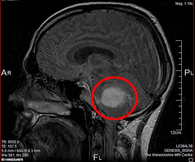
Scientists at the University of California, Davis have built the world’s first combined PET-MRI scanner. Positron Emission Tomography (PET) and the Magnetic Resonance Imaging (MRI), the two kinds of body imaging, have been combined for the first time in a single scanner. Both the imaging modalities are logically complementary, as PET is a functional modality that reports about biological processes, while MRI offers information about tissue structure. The combined scanner can provide accurate PET and MRI images at the same time. It enables the doctors to correlate the structure of a tumour by MRI with the functional information from PET, and to know everything happening inside a tumour. The combined scanners, which are currently available in the market, comprise a combination of Computer-Assisted Tomography (CAT) and PET. MRI scanners depend on strong and smooth magnetic fields that can easily be disturbed by metallic objects inside the scanner. Simultaneously, those magnetic fields can affect the detectors and electronics required for PET scanning. The researchers used a new technology named the silicon avalanche photodiode detector in developing the new combined scanner, as the photomultiplier tubes generally used in conventional PET machines are extremely sensitive to magnetic fields.

 Like
Like
 Dislike
Be a part of Elets Collaborative Initiatives. Join Us for Upcoming Events and explore business opportunities. Like us on Facebook , connect with us on LinkedIn and follow us on Twitter , Instagram.