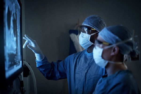

The radiography market has been seeing great innovations of late and with each new innovation there is fresh debate on the pros and cons of the technology vis–vis, ease of use, accessibility and return on investment. The following expert article as well as comments from end users gives us a holistic view of the available technology.

Market Insight Healthcare Practice Frost & Sullivan
Radiography is a method used for the uation of bony structures and soft tissues. An X-ray machine directs electromagnetic radiation, that is, X-rays upon a specified region in the body.
This radiation is capable of penetrating through less dense matter like air, fat, muscle, tissues etc., but is absorbed or scattered by denser materials like bones, tumors and lungs. In film-based radiography, radiation, which passes through the patient, strikes a cassette containing a screen, which has a layer of fluorescent phosphors on it, thus exposing the X-ray film. Areas of the film which get exposed to higher amounts of radiation appear as black or grey on X-ray film depending on the extent of exposure while areas exposed to less radiation will appear lighter or white.

As a medical speciality, radiology can be categorised into Diagnostic Radiology and Therapeutic Radiology. Diagnostic radiology is the interpretation of images of the human body to aid in the diagnosis or prognosis of disease. It is divided into sub fields by anatomic location and application areas, some of which are as follows:
- Chest radiology: It is used for radiological imaging of the chest for detection of diseases.
- Abdominal and Pelvic radiology: This is also referred to as ‘Body Imaging’.
- Interventional radiology: This uses imaging to guide therapeutic and angiographic procedures. At times it is also referred to as Vascular and Interventional radiology.
- Neuro-radiology: It is a sub-specialty in the field of brain, spine, head, and neck imaging.
- Musculoskeletal radiology is the sub-specialty in the field of bone, joint, and muscular imaging.
- Pediatric radiology: used for radiological examination of neonates and children.
Fluoroscopy
Fluoroscopy is the specialised application of X-ray imaging, in which a fluorescent screen or image intensifier tube is connected to a closed-circuit television system, which allows real-time imaging of the anatomical structures in motion with the help of a radio-contrast agent.
The radio-contrast agents are administered, which is usually swallowed or injected into the body of the patient, to delineate the skeletal system, blood vessels and the gastrointestinal tract and renal system. These radio-contrast agents absorb or scatter the radiation, thus allowing real time demonstration of dynamic processes, such as blood flow in arteries and veins. Two radio contrasts are used presently. Barium (as BaSO4) maybe administered orally or through the rectum and is generally used for the uation of the Gastrointestinal (GI) Tract. In some cases, iodine is also used. As Barium is known to cause complications such as tumour, cysts and inflammation. Additionally, in specific situations air can also be used as an agent for the uation of GI system, and carbon dioxide can be used as a contrast agent in the venous system; in these cases, the contrast agent attenuates the X-ray radiation less than the surrounding tissues.
|
Doctor Speak |
|
DDR is quite beyond the reach of individual radiologist in India, it costs in excess of a crore of Rupees, a sum not easily justified in the private sector. Of course the quality of the images are far superior and one would like the luxury of using such technology but cannot afford to. Perhaps select institutions such as the defence, AIIMS or other government hospitals may be able to invest in such technology without thought of returns but not the private sector hospitals. Dr. Rajesh Kapur |
|
We are currently working with CR and digital fluoro radiography (DFR). DFR uses direct images from image intensifiers and is used for interventional and (in our practice) non-interventional procedures like bariums, IVPs etc. This technology is high end and expensive compared to CR systems. DDR of course is the latest and has the highest resolution but is also the most expensive. CR systems are the easiest ones to upgrade and are most portable, apart from being least expensive. DDR is ideal for high throughput hospitals, since that’s the only way to recover investments, and is not viable in low throughput centres. Advantages of DDR include resolution, high throughput, and film-less transfer. Transition and dependence on cross sectional imaging makes CR most viable as a radiography tool. DFR and DDR with/without fluoro is ideal for high throughput hospitals. Dr. Bharat Aggarwal |
Image Digitisation Technology
In radiology, the digital revolution has enabled image enhancement, rapid transmission to remote locations and compact electronic storage. The simplest way to describe the basics of a digital radiography is to relate to the personal digital camera. Earlier people used cameras that had to be loaded with rolls of film, that not only were difficult to load but also cumbersome to manipulate, delete or view images immediately upon capture. The film then had to be developed using chemicals before one could view the images captured. With the introduction of digital technology images could be taken and viewed within seconds, manipulated and have the option of sharing it electronically and to be archived. All this could be done without the use of harmful and expensive chemicals. Given acceptable diagnostic quality, it makes sense to capture images digitally if those images are going to be electronically distributed and stored. The digital image can be printed on film for viewing and/or diagnosis as needed, so a digital imaging system certainly presents an intriguing opportunity to health care providers.
The CR Technology
In Computed Radiography (CR), the X-rays passing through the patient strike a sensitive plate, which is then read and digitised into a computer image by a separate reader. In Digital Radiography the X-rays strike a plate of X-ray sensors producing a digital computer image directly. Plain or analogue radiography was the only imaging modality available during the first 50 years of radiology. It is still the first of the three methods to be used for the uation of the lungs, heart and muskuloskeletal system because of its wide availability, speed, relative low cost and small size which makes it very portable and mobile.
In the last 20 years, CR technology has evolved from an experimental application into a modality with great potential, suited for portable and mobile applications. In the early 1980s, CR products were mostly installed in universities and institutions that laid emphasis on research. The emphasis in the development of CR technology today revolves around the size of the hardware and the diagnostic quality of the images. By the late 1980s, commercial CR products were available and installed in hospitals where radiologists were keen to learn this new technology which had better image quality and resolution in comparison to the old film based system. During its technological development over the last years, the CR hardware has become more and more compact.
Today, the computed radiography system is seen as an effective and efficient method of delivering radiographic images in critical situations where conditions are difficult for radiologists to obtain consistent images on radiographic film. In such situations, CR systems are capable of generating images with excellent diagnostic value and superior resolution. CR systems are capable of growing further because the radiation dose used is much less when compared to the dosage used for screen or film techniques.
Improvements in storage phosphor materials, image processing software, optical collecting systems and laser scanners combine to boost the sensitivity of modern CR systems. Some manufacturers are in their fourth or fifth generation of development.
Image Quality of CR Systems
While the radiation dose of CR systems is comparable to film-based systems, spatial resolution of the same is not comparable. The spatial resolution of a general screen/film system is 7 to 8 line pairs per millimeter whereas that of a CR system is less than half that, ranging from 2.5 to 5 line pairs per millimeter. In majority of the applications, CR does not perform well in terms of spatial resolution, but has a very high contrast resolution, which enhances the diagnostic utility of the images.
The CR systems are equipped with the ability to adjust levels of brightness in images, which gives radiologists the ability to view structures that cannot be easily detected on radiographic film. With film, the contrast is determined by the composition of the film, the radiographic technique and the chemical processing. The main advantage of computed radiography is noted in it being portable and mobile, where the CR characteristics reduce the number of repeat exams required due to inaccurate technique adjustment or exposure. CR also provides the ability to provide rapid electronic image distribution.
Direct Digital Radiography (DDR) Technology
The direct digital radiography technology uses a direct process to convert X-ray energy to digital signal. The image is directly captured on the flat plate and is then viewed in the computer. This technology uses amorphous selenium (a-Se) or an amorphous silicon (a-Si) flat plate placed in between the object to be diagnosed just as the process carried out for film-based systems.
A number of components are required for direct digital image production, which includes an X-ray source, an electronic sensor, a digital interface card, a computer with an analog-to-digital converter (ADC), screen monitor software and a printer. Direct digital sensors are either a charge-coupled device (CCD) or complementary metal oxide semiconductor active pixel sensor (CMOS-APS). CCD is a solid-state detector composed of an array of X-ray sensitive pixels on a pure silicon chip. Charge coupling is a process where the number of electrons deposited in each pixel is transferred from one to another in a sequence manner so as to obtain an amplified image output on the monitor. Fiber optically coupled sensors utilise a scintillation screen coupled to a CCD. The complementary metal oxide semiconductor active pixel sensor (CMOS-APS) is the latest development in direct digital sensor technology. The CMOS sensors are identical to CCD detectors but they use an active pixel technology and are less expensive to manufacture having the same image quality as that of the CCD detectors.
The Future of Radiology
 Dramatic changes are expected in radiology in the coming years, including widespread use of digital technologies. Radiology departments may apply screen/film, CR and digital radiography systems to various applications, depending on the requirement of the departments. Thin-film transistor digital radiography technology is one of the evolving technologies for radiology. In this technology, an array of transistors converts X-ray energy to an image. Electronic scanning of the transistors collects data. Unlike CR, phosphors or X-ray converting overcoats may or may not be involved, and there is no need for a laser to scan the imaging plane. This technology will have the advantage because technologists no longer will have to carry the cassettes to a processing station.
Charged coupled device (CCD) technology is also an alternative for direct digital capture.
Over the long term, digital radiography will likely evolve as stand-alone system for chest imaging and digital X-ray tables. Along with this, CR systems will continue to be in use for mobile X-ray applications as customers seek avenues to create the-all-digital department.
The ability to digitise a film image and transmit the data to remote sites will continue to aide the CR and DR technologies to further evolve. 
Be a part of Elets Collaborative Initiatives. Join Us for Upcoming Events and explore business opportunities. Like us on Facebook , connect with us on LinkedIn and follow us on Twitter , Instagram.


