Article
PET CT – An advanced, Innovative Tool to Detect Cancer
PET CT – An advanced, Innovative Tool to Detect Cancer
![]() By Elets News Network - 25 April 2019
By Elets News Network - 25 April 2019


PET (Positron Emission Tomography) scan is a nuclear medicine technique, which produces images at a molecular level. It is a whole body scan, where radiopharmaceuticals or “tracers” labelled with radioactive isotopes of carbon, oxygen, nitrogen and fluorine are used.
These isotopes mimic proteins, oxygen, water and glucose. Most commonly used radio-isotope in clinical practice is fluorine-18 (18F) labeled fluoro-deoxy-glucose (FDG).
These radioisotopes act like molecular level probes which target disease while giving out radio-signals that are captured by ultrasensitive detectors to produce an image.
PET scan thus reveals much more about the cellular level dynamics of a disease than any other modalities like ultrasound, CT scan or MRI. With combination of CT / MRI with PET into a single machine, simultaneous localisation of the disease becomes more accurate with better characterisation of individual lesion(s).

The major areas where PET is making critical contributions include cancer care, coronary artery disease (heart attack), neurology and to some extent in psychiatry.

ONCOLOGY
PET scans are helpful in distinguishing benign (non-cancerous) from malignant (cancerous) disease when carried out at initial stage of diagnosis of cancer. The technique also helps to determine its stage.
The metabolic characteristics of the cancer help physicians to decide the best treatment options for such patients. Subsequently, during the course of treatment the PET scans show how the cancer is responding to the treatment.
As the cancer responds to treatment, the metabolic activity of tumour, as seen in PET scan, starts diminishing which proves that the therapy / treatment is working.
On the other hand, if there is no response or if cancer is growing despite treatment, an early PET scan can alert treating physician to alter the treatment protocol thus sparing patient the unnecessary and often very expensive and painful treatment.
NEUROLOGY
PET scans can diagnose Alzheimer’s disease if early intervention is made. It can also locate tumour in brain and distinguish active viable tumour from post radiation scar tissue. In certain cases of epilepsy, PET scans are used to locate the focus of seizures and accurately assess sites within brain for delicate surgery.
CARDIOLOGY
In case of heart attack (myocardial infarction, acute coronary syndrome), a PET scan helps to determine myocardial viability. It gives idea whether coronary bypass surgery (CABG) is required or not and what benefits it would bring.
PSYCHIATRY
Schizophrenia, depression, dementia and substance abuse are some of the indications for PET scan, which provides new insights into the biology of the disease, enabling doctors to plan better management protocol and close follow-up.
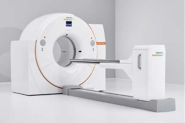
The “Nuclear Medicine & Molecular Imaging department” at Sarvodaya Hospital and Research Center, Faridabad, has recently commissioned a brand new PET-CT scanner from GE (Discovery IQ). It is equipped with state-of-the-art 50 slices per second CT. The machine uses Q-Clear reconstruction algorithm which is the latest innovation in PET technology.
The technology helps the machine to detect the tiniest lesions, thus increasing the accuracy of the investigation. The machine makes entire procedure safe and more comfortable for patients.
(Spl. Note: The writer, Dr Swagat Dash, is a Senior Consultant and HoD – Nuclear Medicine, Sarvodaya Hospital & Research Centre, Faridabad. Views expressed are a personal opinion.)

 Like
Like
 Dislike
Be a part of Elets Collaborative Initiatives. Join Us for Upcoming Events and explore business opportunities. Like us on Facebook , connect with us on LinkedIn and follow us on Twitter , Instagram.
Disclaimer: The views and opinions expressed in this article are solely those of the author and do not necessarily reflect the official policy or views of any organisation. The content is intended for informational and educational purposes only and should not be construed as medical advice.
"Exciting news! Elets technomedia is now on WhatsApp Channels Subscribe today by clicking the link and stay updated with the latest insights!" Click here!