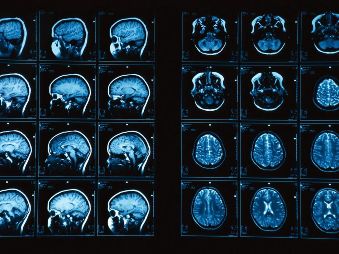
New research shows that the addition of two magnetic resonance imaging (MRI) sequences to a common MR angiography technique significantly improves detection of pulmonary embolism, a potentially life-threatening condition traditionally diagnosed through computed tomography (CT). Results of the study are published online in the journal Radiology.
Pulmonary embolism occurs when a blood clot”usually from the leg”travels to the lung and blocks the pulmonary artery or one of its main branches. CT angiography is the gold standard for diagnosis, but it exposes patients to ionizing radiation and iodinated contrast agent, which carries a risk of allergic reactions and kidney damage in some patients.
“MRI is developing much faster than CT,” said Diego R. Martin, M.D., Ph.D., head of the Department of Radiology at the University of Arizona College of Medicine in Tucson. “The images we’re getting are already significantly better than they were a year ago. There is no doubt that in the future we will be able to offer a non-radiation-based alternative to CT for the diagnosis of pulmonary embolism.”
MRI has been used for pulmonary embolism detection in pregnant women and patients whose kidneys may be harmed by CT angiography contrast agents. However, the Prospective Investigation of Pulmonary Embolism Diagnosis III (PIOPED III) study, a major multicenter trial, found that centers had difficulty obtaining adequate quality MR pulmonary angiography (MRPA) for suspected pulmonary embolism. The PIOPED researchers determined that MRPA should be considered only at centers that routinely perform it and perform it well, and for patients who have contraindications to standard tests.
In the new study, the research team assessed the impact of two additional MRI sequences on MRPA’s accuracy. The two techniques”contrast-enhanced volumetric interpolated breath-hold examination (VIBE) and non-contrast true fast imaging with steady-state precession (true FISP)”complement MRPA, according to Dr. Martin.
The addition of VIBE provides a gray scale that enables readers to distinguish between the clot, or thrombus, and the lung, which both appear dark on MRPA.
The true FISP test does not require contrast agent or a breath hold, an important consideration for embolism patients who often cannot hold their breath long enough for image acquisition on MRPA.
When Dr Martin and colleagues studied the three techniques on 22 patients with CTA diagnosis of pulmonary embolism, they found a sensitivity of 55 percent, 67 percent and 73 percent for MRPA, true FISP and VIBE, respectively. Combining all three MRI sequences improved the overall detection rate to 84 percent. Specificity was 100 percent for all detection methods except for MRPA, which demonstrated one false positive.


 Like
Like
 Dislike
Be a part of Elets Collaborative Initiatives. Join Us for Upcoming Events and explore business opportunities. Like us on Facebook , connect with us on LinkedIn and follow us on Twitter , Instagram.
"Exciting news! Elets technomedia is now on WhatsApp Channels Subscribe today by clicking the link and stay updated with the latest insights!" Click here!