


Research
3D X-Rays in Orthodontic Diagnosis Increase Radiation Exposure
3D X-Rays in Orthodontic Diagnosis Increase Radiation Exposure
![]() By eHealth Network - 03 February 2011
By eHealth Network - 03 February 2011


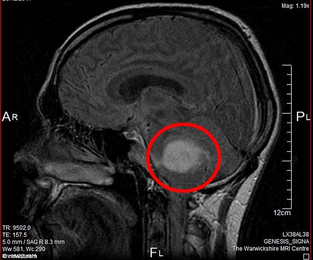
The use of 3D X-rays in orthodontic cases may expose patients to unnecessary radiation that could be avoided by using 2D X-rays, a study published in Dentomaxillofacial Radiology has warned. The study found that one image of 3D cone-beam CT produces around 87-200mSv of radiation, compared to 4-40mSv for an entire series of 2D X-rays required for orthodontic diagnosis. Study author Dr Erika Benavides said that adding unnecessary radiation exposure to the patient may result in higher biological risks, particularly in young children. “This is why selecting the patients that would benefit the most from this additional exposure needs to be done on a case-by-case basis,” Benavides added. The researchers noted that when used judiciously, cone-beam CT is an invaluable tool with a definite place in orthodontic treatment planning.


 Like
Like
 Dislike
Be a part of Elets Collaborative Initiatives. Join Us for Upcoming Events and explore business opportunities. Like us on Facebook , connect with us on LinkedIn and follow us on Twitter , Instagram.
"Exciting news! Elets technomedia is now on WhatsApp Channels Subscribe today by clicking the link and stay updated with the latest insights!" Click here!