April 2014
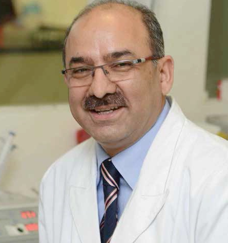
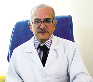
Today toddlers Bounce Back from CHD – Dr Vikas Kohli, Director & HOD, Pediatric Cardiology, BLK Super Speciality Hospital
Today toddlers Bounce Back from CHD – Dr Vikas Kohli, Director & HOD, Pediatric Cardiology, BLK Super Speciality Hospital
![]() By eHealth Network - 11 April 2014
By eHealth Network - 11 April 2014


 Recent advances in medicine ensures that children born with a heart defect live healthy lives. Dr Vikas Kohli, Director & HOD, Pediatric Cardiology, BLK Super Speciality Hospital, in conversation with Shahid Akhter, ENN, focuses on the issue of Congenital Heart Disease (CHD)
Please tell us about the congenital heart defects (CHD) today?

The commonest birth defect seen in infants and newborns today are related to the heart. The abnormality is seen in almost 1 in 100 pregnancies. Congenital heart defects commences in the early stages of pregnancy, when the heart is developing. This includes abnormalities in the hearts structure, electrical system and other abnormalities that affect the function of a babys heart.
Usually it is genetic or chromosomal abnormalities that lead to heart defects. Risk factors may include smoking during pregnancy, maternal obesity, maternal diabetes and maternal infections such as rubella.
Congenital heart disease is a general term for a wide range of birth defects that affect the normal workings of the heart. We are not too sure about the causes leading to congenital heart disease. Some know risk factors may include.
* Downs syndrome “ a genetic disorder that affects a babys normal physical development and causes learning difficulties.
* Mother having certain infections, such as rubella, during pregnancy
* Mother having poorly controlled type 1 diabetes or type 2 diabetes.
What is a Heart Murmur ?
After birth, the diagnosis usually involves a pediatric doctor hearing a heart murmur. A heart murmur happens to be an abnormal heart sound. Once this is suspected, a cardiologist performs an echocardiogram and a confirmation of whether the murmur is from an abnormality in the heart or is an innocent murmur can be made. Innocent murmur is a murmur which though present is not associated with a heart abnormality i.e. the heart is innocent and normal though there is a murmur. This is a fairly common situation.
How do you classify the heart diseases ?
There are 2 main types of heart disease: one in which the baby turns blue and the ones in which the baby does not turn blue.
Blue Baby
Almost all conditions in which the baby turns blue require surgical treatment. For others there may at times not be a requirement for surgery or it may be treatable by balloon angioplasty or device closure. Both these methods are non-surgical methods. All major defects require surgery to be done.
Hole in the Heart
The more common defect of the heart involves a hole in the heart. An isolated hole in the heart (which could be a ventricular or atrial defect), will require treatment. The hole is between chambers carrying red and blue blood (red signifying with oxygen and blue signifying without oxygen). The condition of isolated hole in heart should not be confused with conditions where hole is present associated with many other abnormalities in the heart, which have varying treatment and outlook.
What forms of treatments are available ?
Many conditions now can be treated without surgery. The closure of such defects can be done by an angioplasty technique similar to the one in adults to place stents.
The common problems in children include a hole between the lower chambers (called VSD). The wall between the upper (ASD) and lower (VSD) chambers separates the red from the blue blood. A hole would result in extra blood flow to the lungs. This makes the child have more chest infections, child gains weight with difficulty and feeding also becomes a problem.
On the other hand, the child could be blue when in addition to a hole in heart, there is a blockage of blood flow to the lungs (ToF). This is the commonest condition in which the baby becomes blue. Such conditions always require surgery to be done. Other defects in which the child becomes blue includes where the red blood from lung (with oxygen) drains abnormally into the blue blood, or the tubes coming out of the heart carrying red and blue blood get switched whereby the body receives blue blood wrongly and the lungs get red blood. These conditions usually require a single operation and the child becomes normal. Finally, it is the condition when one of the valves or of the pump is not normal that the child requires more than one operation in lifetime of a patient and may affect the quality of life or the life span.
How Early Can the diagnosis be made ?
With the current technology the diagnosis can be made of heart disease in the 18th week of pregnancy. This test is called fetal echocardiogram. Specialised heart ultrasound machines are used for this test with advanced features like STIC or FETAL NAVIGATION. Once the diagnosis is made the family can be counselled for future treatment. Or if the condition requires complicated treatment, the pregnancy can be terminated too.
If an abnormality is noted, often it is a condition of the heart which does not require immediate treatment or surgery. Occasionally the abnormality may warrant intervention or surgery”
What is Device Closure of a hole in heart ?
This technique involves implantation of one of several devices (basically single or double wire frames covered by fabric) using heart catheterization methods in the cardiac catheterization laboratory, without the need for cardiopulmonary bypass (heart-lung machine), and without the need to stop the heart. Defects amenable to such device therapy tend to be smaller (less than 20 to 25 mm [3/4 to 1 inch] diameter). Importantly, these lesions must be centrally located within the atrial septum. Defects at the very upper or lower edges of the atrial septum (called ostiumprimum or sinus venosus) are not good candidates for this procedure, because these defects usually involve other abnormalities of the heart valves, or venous drainage from the lungs. This determination can be made by the patients primary cardiologist.
The usual procedure is very similar to standard heart catheterization. Briefly, flexible long tubes (or catheters) are inserted into the veins and arteries in the groin. We use the knowledge that in all human beings, these vessels are directly attached to the heart, and this is the standard access technique used in all patients. Routine pressures and oxygen levels in all of the chambers of the heart are then obtained. Angiograms (pictures taken following dye injection) are performed to determine the size of the chambers, the size of the defect, and its location within the heart. Using a balloon catheter of a known diameter, the defect is then sized in comparison to the balloon, so that the device appropriate for that particular patient can be chosen. The device is then advanced into the heart through an introducer sheath (larger, less flexible tube).
- Congenital heart disease (CHD) causes the death of thousands of children in developing countries each year. Eight out of every 1000 live births around the world have some form of CHD.
- Each year, approximately 1 million babies are born with heart defects worldwide.
 Like
Like
 Dislike
Be a part of Elets Collaborative Initiatives. Join Us for Upcoming Events and explore business opportunities. Like us on Facebook , connect with us on LinkedIn and follow us on Twitter , Instagram.