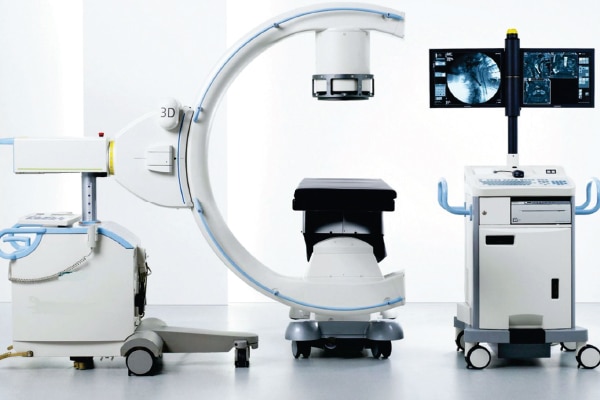
The modern-day advancement has also influenced radiology. About 20 years ago, radiology was limited to X-rays. But over the years, conventional procedures have witnessed tremendous change for the better, leading to enhanced experience of treatment for millions of people across the globe.
Of the most recent achievements, the prominent ones can be mentioned as — Digital Radiography and Digital Mammography, Ultrasonography, Remote viewing system, CT Angiography, Replacement of Exploratory Surgery, etc.

Digital Radiography and Digital Mammography

 Digital radiography has tremendously improved the X-ray quality, speed and replaced all the boring darkroom procedures, as these can be shared immediately for speedy diagnostic results. The conventional X-ray used to take about half-an-hour for whole procedure while the digital radiograph can be acquired with just a click of a button while delivering far better quality.

USG: Ultrasonography works on the principle of getting echo from reflected sound waves. It has advanced tremendously over the years from 2D to 4D images and remote viewing systems of the present day. USG is very important imaging modality. It’s different from other imaging modality as it is real time and safer, as it does not involve radiations. USG has also advanced over the years with dopplers, guided needle placement elastography and many more.

Remote viewing system
Radiology has advanced immensely over the years but the sad reality is that in India and many other countries a lot of people don’t have access to basic radiological procedures.
Kenya with a population of 43 million has only 200 radiologists. In rural Nepal, there are places where people travel almost two days for getting X-ray. In that part of the world, machines are often outdated models and are the ones that have been donated.
Web-based systems allow physicians to send and access images and reports from all over the world.
CT Angiography
Traditional angiography takes several hours, requires sedatives and can even cause damage to vessels. But CT- Angiography takes 10-15 minutes without such risks. It can be used to image any artery in the body including brain, coronary, lung, kidney etc. with recent advances in CT angiography. It gives a picture which is three-dimensional and is like actual arterial system in our body and is very friendly for clinicians to see the pathology.
Replacement of Exploratory surgery
CT scan reduces negative appendectomy. It is very important in trauma patient where decision has to be taken from CT irrespective of the patient needs surgery or not. So reducing trauma caused by surgery and saving a lot of patients’ expenditure done on unnecessary surgeries.
MRI
MRI plays a crucial role in detecting pathology at its earliest. In stroke patients, it can detect the changes seen in Acute infarcts (a small localised area of dead tissue resulting from failure of blood supply) at its earliest.
Recent advances in MRI like spectroscopy magnetisation transfer and functional MRI plays a crucial role in deciding the management and type of therapy. MRI is radiationless and gives very accurate and minute details of pathology.
PET – CT
Positron emission tomography continued with CT scan images provides easy detection of even small tumours and metastatic information and also provide metabolic status of tumour as physicians get a better idea of what is going on and how to properly treat it and also help in monitoring chemotherapy.
In brief, Radiodiagnosis has tremendously improved over the previous years. New technologies and better practices have made the field safer, less expensive and more efficient.
Be a part of Elets Collaborative Initiatives. Join Us for Upcoming Events and explore business opportunities. Like us on Facebook , connect with us on LinkedIn and follow us on Twitter , Instagram.
"Exciting news! Elets technomedia is now on WhatsApp Channels Subscribe today by clicking the link and stay updated with the latest insights!" Click here!


