 Medical technology is undoubtedly indispensable to the health and for improving quality of life of people. It has revolutionised healthcare from almost past three decades, allowing doctors to find disease earlier and improve patient outcomes.
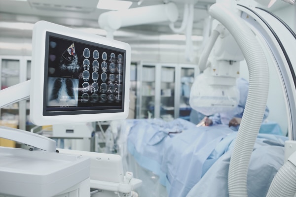
In a utopian world, it would have been possible to diagnose, treat and cure patients without causing any harmful side-effects. Since this is not possible today, the efficacy of medical imaging cannot be overstated considering it has enabled doctors to see inside a patient without having to cut them open.

It has ensured early detection and treatment for cases such as lung, breast cancers etc. The chances of medico-legal issues too have been reduced due to delayed or incorrect diagnosis.
There is some downside linked with the imaging technology. In the present era, the deterioration of skills during physical examination of patients has become much more evident.
Conventional ultrasound imaging relies on good contact between the transducer and the skin, using acoustic coupling gel. Without this, the ultrasound beam encounters a large acoustic impedance mismatch between the transducer and the air in front of it, which prevents beam transmission. This limits the use of ultrasound in some clinical scenarios, including imaging of open wounds and during surgery, where sterile conditions are required.
Now, researchers in Japan have demonstrated non-contact ultrasound imaging, where the transducers are separated from the skin by air. “The project is in its infancy, but the results to date have been very encouraging,” claims Gregory Clement, physicist and lead author of a study conducted at the University of Electro- Communications in Tokyo.
The non-contact technique exploits the greater focusing potential of ultrasound in air. A high beam intensity in air compensates for the low transmission of the beam across the air–skin boundary.
The group’s approach uses a bowl-shaped transducer to focus an ultrasound beam on to a 5 mm spot at the skin surface. The spot generates a point source of ultrasound that when scanned over the surface of an object, can be used to perform diffraction tomography.“The method treats the skin surface as a ‘virtual’ transducer, allowing for a large effective number of sources and receivers, thus opening the potential for large regions to be imaged,” claims Clement.
“Current medical probes can’t conform to the body, thereby limiting where they can be placed.” The team simulated and constructed a 40 kHz transducer prototype, comprising 400 elements arranged in a hemispherical array.
After beam characterization and transmission measurements, they acquired images using the transducer and a broadband microphone receiver.
In vivo, non-contact imaging of a hand was demonstrated with a conventional C-mode approach, which produces images for a fixed depth below the skin. The low- range scan showed varying contrast between the anatomy containing mainly bone and that containing only soft tissue.
Clement says that the group will continue the development and clinical testing of the non-contact imaging technique over the next few years. Ongoing work in the lab includes optimising the receiver to increase technique sensitivity and investigating faster acquisition techniques to reduce scan times.
The ability to visualise processes that take place in the brain during the development and progression of Alzheimer’s disease also provides a powerful aid for diagnosing condition, monitoring treatments and testing preventive and therapeutic agents.
With this aim, a research team at the National Institute of Radiological Sciences in Japan has developed a new type of imaging agent that enables visualisation of “tau-protein” within the brain of living patients.
In patients with Alzheimer’s disease, tau proteins aggregate together and become tangled, while fragments of another protein, amyloid beta, accumulate into plaques. Tau tangles are not only an important marker of neuro-degeneration in Alzheimer’s disease, but also a hallmark of other neuro-degenerative disorders that do not involve amyloid-beta plaques. While imaging technologies have been developed to observe spread of amyloid-beta plaques within patients’ brain, tau tangles have previously not been easily monitored in living patients.
The researchers have now developed fluorescent compounds called PBBs that bind to tau inclusions and can be visualised using positron emission tomography (PET) and optical imaging. They showed that the PBBs could detect tau lesions in a mouse and a living patient. In the clinical PETstudy, PBB revealed a strong signal in the hippocampus of a patient with Alzheimer’s disease, in contrast to PET images recorded usinga beta-plaque tracer. “PET images of tau accumulation are highly complementary to images of senile amyloid-beta plaques,” claims the Institute. This is of critical significance because tau lesions are known to be more intimately associated with neuronal loss than senile plaques.
There are some other latest cutting-edge imaging techniques that may become popular in the near future. Pocket-sized hand-held ultrasound devices are predicted to replace the 200-year-old stethoscopes in near future. They can diagnose heart, lung and other problems more accurately than traditional stethoscopes.
Similarly, Hyperspectral imaging may provide a non-invasive diagnostic method that allows determination of pathological tissue with high reliability. There are chances that this would also lessen doctor’s liability and reduce indemnity insurance premiums. This technology is in use in the defence sector and is now finding applications in the healthcare imaging Industry.
A modality called Electromagnetic Acoustic Imaging developed by the University of Oxford has been making use of electro-magnetic and acoustic waves for diagnosis. It helps to identify various types of solid Cancers in the early stages of development. Another technology that goes by the name Wafer Scale Mega Microchip developed by the University of Lincolnis also used to enhance medical imaging techniques. The University says that images that are produced by the chip will enable doctors to detect accurately the effects of radiation on cancerous tumours, thus helping early detection.
Besides, there are two other universities – the University of California, Berkeley and the Universidad Autonoma de Madrid of Spain who have claimed to have made the use of 3D Meta material to augment ultrasound images by a factor of 50X. If this technology becomes successful commercially, it would help current ultrasound investigations to capture high- resolution images for medical imaging in both diagnosis and interventional procedures.
Another claim is by the University of Medicine, Berlin and Max- Delbruck Centre for Molecular Medicine, Berlin, who claims that they have produced a technology for capturing images of the beating heart in its MRI systems. They claim the magnetic field of resolution would be 1, 50,000 times the earth’s magnetic field. It would thus aid in clearly demarcating between blood and heart muscle, enabling early diagnosis of cardiac malfunction.
Scientists at Japan’s Railway Technical Research Institute, Tokyo, too have managed to develop a palm-size superconducting magnetic system. This would make current MRI System into mobile imaging applications.
Iron oxide nano crystal technology is being considered for use in medical imaging. Super paramagnetic iron oxide particles have a variety of applications in molecular and cellular imaging. Evaluation of some of the recent research has concerned cellular imaging with imaging of in vivo macrophage activity. As per the iron oxide nano particle composition and size which influence their bio-distribution, several clinical applications are possible. The use can be checked for detection of liver metastases in cancers, metastatic lymph nodes, inflammatory and degenerative diseases.
Research is also on in the field of stem cell migration and immune cell trafficking, as well as in the areas of targeted iron oxide nano particles for molecular imaging studies.
Another study that is under focus is that on the ultra small super paramagnetic iron oxide particles that are being researched as blood pooling agents for angiography, tumour permeability and tumour blood volume or steady-state cerebral blood volume and blood vessel size index measurements.
3D bio-printing has undergone seminal advancements in recent times. 3D bio printing is the process of creating cell patterns in a confined space using 3D printing technologies, where cell function and viability are preserved within the printed construct. This technique allows high precision fabrication of biological structures encapsulating cells and bioactive molecules, with applications in areas such as tissue engineering, drug development and bio-sensing.
Recent advances have enabled 3D printing of biocompatible materials, cells and supporting components into complex 3D functional living tissues. 3D bio printing is being applied to regenerative medicine to address the need for tissues and organs suitable for transplantation. Compared with non-biological printing, 3D bio printing involves additional complexities, such as the choice of materials, cell types, growth and differentiation factors, and technical challenges related to the sensitivities of living cells and the construction of tissues.
3D bio printing has already been used for the generation and transplantation of several tissues, including multilayered skin, bone, vascular grafts, tracheal splints, heart tissue and cartilaginous structures. Other applications include developing 3D-bioprinted tissue models for research, drug discovery and toxicology.
Researchers at Siemens Corporate Technology have developed learning- based software that can identify and section out any organ in any digital medical image, irrespective of occlusions, angle of view, imaging modality, or pathology.
The company says that this method can be used to synthesise a virtual high dose CT image from a low dose CT image, enabling reduction of the radiation dose to which the patient is exposed. Siemens has reportedly applied for a patent.
The John Hopkins University has developed a procedure for precise characterisation of tumour burden and response to therapy from PET Scans. Although this method is awaiting patent approval, it is known to provide reliable and reproducible PET tumour boundary definition and metabolic tumour volumetric quantification over a wide range of tumour sizes and shapes and for various levels of PET radiotracer uptake.
This method will help clinicians in diagnosis, staging and planning of radiotherapy and surgery as well as determining treatment response. It can also help in cancer therapy research.
At the Ohio State University, researchers have studied a new method for performing CT-based elastography. This method estimates local displacement of tissue in response to a mechanical stimulus from high- resolution images. It can also be applied to hard structures in the body like bone, teeth and cartilage as well as soft tissues. It can also be used to scan patients with pace- makers or metal implants that cannot undergo MR scans.
A new technique has been developed by researchers at Philips that determines blood flow velocity. Philips has applied a patent for it. The method uses ultrasound pulse at one location and the pulse is detected at a second location by an ultrasound receiver. The calculation is then done for flow velocity of blood in a blood vessel between the first and second locations.
Barbados-based Synaptive Medical Company has developed a new technique for correcting warping in MR images caused by non-linearities in gradient field profiles. The patent for this new technique is pending.
With so many advancements in technology, healthcare delivery has witnessed a tangible progress in terms of patient care. Nonetheless, use of high-end technologies has escalated the cost of quality healthcare facility. A section of people finds it tough to avail healthcare facility at increased price. High-out-of- pocket expenditure has become a big concern.
The need of the hour is to calibrate both these factors i.e affordability and costly technologies, to ensure best healthcare facilities to the people at large.
 Like
Like
 Dislike
Be a part of Elets Collaborative Initiatives. Join Us for Upcoming Events and explore business opportunities. Like us on Facebook , connect with us on LinkedIn and follow us on Twitter , Instagram.