Genomics
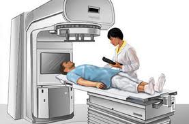
Combining oxygen with radiation therapy decelerates many cancer tumors
Combining oxygen with radiation therapy decelerates many cancer tumors
![]() By eHealth Network - 29 July 2013
By eHealth Network - 29 July 2013


A multidisciplinary team at UT Southwestern Medical Center has found that measuring the oxygenation of tumors can be a valuable tool in guiding radiation therapy, opening the door for personalized therapies that keep tumors in check with oxygen enhancement.

In research examining tissue oxygenation levels and predicting radiation response, UT Southwestern scientists led by Dr. Ralph Mason reported in a recent online issue of Magnetic Resonance in Medicine that countering hypoxic and aggressive tumors with an “oxygen challenge” – inhaling oxygen while monitoring tumor response – coincides with a greater delay in tumor growth in an irradiated animal model.
Over the past several years, the research of Dr. Mason, professor of radiology and the paper’s senior author, and his colleagues has been building on findings that show lack of oxygen actually stimulates the growth of new blood vessels in tumors and leads to metastasis and genetic instability in cancer. The theory follows that breathing oxygen or enriching the oxygen content of hypoxic (low in oxygen) cancer tissues improves therapy.
In the current study, supported by the National Cancer Institute, smaller tumors based on magnetic resonance imaging were found to be significantly better oxygenated than larger ones. This confirmed previous investigations that show a range of hypoxic environments depending on the size of the tumor.
“The next step is clinical trials to assess tumor response to radiation therapy,” said Dr. Mason, director of the cancer imaging program at the medical center. “Tumors determined to be hypoxic can be uated and made responsive through mild and easy-to-administer interventions, such as breathing more oxygen or taking a vasoactive drug. Monitoring the response to oxygen breathing tells us which tumors will benefit.”
If the results are confirmed in humans, the implications for personalized therapies for other cancers could mean fewer radiation treatments, or perhaps, ideally, one single high-dose treatment. Lung cancer, for instance, is a form of the disease whose tumors are poorly oxygenated despite being located in the principle organ charged with oxygenating the blood.
“The ability to stratify tumors based on hypoxia offers new opportunities to tailor therapy to tumor characteristics, potentially enhancing success through personalized medicine,” Dr. Mason said.
Together with Dr. Robert Timmerman, professor of radiation oncology at the Harold C. Simmons Cancer Center, and Dr. Ivan Pedrosa, professor of radiology and the Advanced Imaging Research Center, Dr. Mason is starting clinical trials to assess the effectiveness of oxygenation during treatment with stereotactic body radiation in humans – work that is supported by the Cancer Prevention and Research Institute of Texas (CPRIT) through one of its Multi-Investigator Research Awards.
With CPRIT support, Dr. Mason’s team has worked to understand how low oxygen concentration can cause radiation resistance in tumors. In some cases, the simple addition of oxygen to stereotactic body radiation greatly improves response. The key is to identify those patients who will benefit.
 Like
Like
 Dislike
Be a part of Elets Collaborative Initiatives. Join Us for Upcoming Events and explore business opportunities. Like us on Facebook , connect with us on LinkedIn and follow us on Twitter , Instagram.