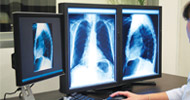
 A workstation without tools will be like a soldier sans his armour radiology has not been untouched by the rapid advances in technology. Our radiologists are able to serve the needs of many people by using the high-tech tools in their workstation
By Dr Amar Jain

works as a clinical radiologist for HealthFore “ Religare Technologies. He is an IT evangelist who is working with the aim of syncing technology and diagnostic radiology. He can be contacted @amar.jain@religaretech.com.
A zoom tool enlarges the entire image and then one needs to pan to the site of interest, whereas eth magnifying tool will only magnify the region of interest. There are also orientation tools like flip tools, rotate and invert tools to enable us to carry on the examination in whichever position we desire. We can also flip, rotate or invert in the desired position of view.
The bookmark tool helps us to store the annotations (measurements) applied to the image and retrieve it back as and when required. A radiologist, who reads a particular image highlights the abnormality, marks the measurements and bookmarks it. All modalities have a ruler, line, text, free hand tools as basic tools to measure standardised length, breadth and depth and highlight their specifications. The layout tool compares different studies, series done at the same time or over a period of time.
Hanging protocol allows consistent viewing of images based on body parts and modality type. As the word explains, this tool hangs the images on different monitors rather than view boxes, as used to happen with developed films. While training over 60 radiologists during implementation of RIS-PACS by HealthFore implementation team, a number of questions came up, regarding the tools and their clinical relevance.
Probe showing h.u. Value is a tool found in modalities such as plain CT images, contrast enhanced, CT images, HRCT images and CT angiography images. It is used to differentiate blood from dystrophic calcification, to assess the different stages of bleed if its acute or chronic and to see h.u. Value changes before and after contrast injection to uate mass lesions. Such modalities also have tools like window width and level tool to select appropriate window for appropriate study. There is mediastinal window to study bronchogenic CA and its mediastinal extensions for staging. CT images, MRI images, CR and DR images, DSA images endoscopic and virtual bronchscopic images have tools such as zoom and pan to see a nidus and feeder artery in an arteriovenous malformation and detect aneurysms. Cline tool helps see all images in a video mode by setting appropriate speed. Coronary analysis tool for cardiac CT provides a number of multi-planar reformat (MPR) views. The 3di PET/CT viewer contains different viewing sections for the PET and CT, MPR images, and for the fused MPR images of each series. 3di brain perfusion feature highlights optimised colour display of results maps for CBV (cerebral blood volume), CBF (cerebral blood flow), MTT (mean transition time) and TTP (time to peak). These maps provide invaluable clinical information to assist clinicians in stroke assessment and treatment planning.
 About Author
Dr Amar Jain
works as a clinical radiologist for HealthFore “ Religare Technologies. He is an IT evangelist who is working with the aim of syncing technology and diagnostic radiology. He can be contacted @amar.jain@religaretech.com..
 Like
Like
 Dislike
Be a part of Elets Collaborative Initiatives. Join Us for Upcoming Events and explore business opportunities. Like us on Facebook , connect with us on LinkedIn and follow us on Twitter , Instagram.